Lower Body Diagram / Lower Body Diagrams Lzr Ultrabright Powerful Led Therapy / Related posts of anatomy of lower body anatomical drawings.
byAdmin•
0
Lower Body Diagram / Lower Body Diagrams Lzr Ultrabright Powerful Led Therapy / Related posts of anatomy of lower body anatomical drawings.. These muscles provide posture and stability to the body by holding the vertebral column erect and adjusting the position of the body to maintain balance. B) free body diagram of point p; At the level of the pelvic bones, the abdomen. The knee joins the upper leg and the lower leg. The lower, narrow part of the uterus (womb) located between the bladder and the rectum.
A free body diagram is a graphic, dematerialized, symbolic representation of the body (structure, element or segment of an element) in which all connecting pieces have been removed. 12 photos of the muscles of the lower back and buttocks diagram. Evenly distribute weights from your upper body into the lower extremities. We hope this picture human body artery diagram in detail can help you study and research. In all, there are believed to be 80 organs in your body, all serving different functions and uses.
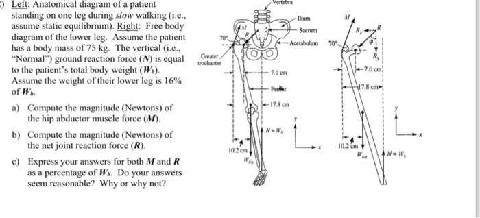
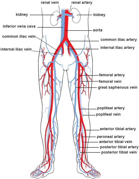
Left Anatomical Diagram Of A Patient Standing On One Chegg Com from d2vlcm61l7u1fs.cloudfront.net The iliac, femoral, popliteal and tibial (calf) veins are the deep veins in the legs. The back functions are many, such as to house and protect the spinal cord , hold the body and head upright, and adjust the movements of the upper and lower limbs. A diagram shows the various inguinal lymph nodes (lymphatic ganglia). Sensory nerves are nerves that. Anatomynote.com found human body artery diagram in detail from plenty of anatomical pictures on the internet. Two forces (lower part of figure below) 1) the weight w exerted by the earth on the box. Learn vocabulary, terms, and more with flashcards, games, and other study tools. The lower, narrow part of the uterus (womb) located between the bladder and the rectum.
A free body diagram is a graphic, dematerialized, symbolic representation.
B) free body diagram of point p; Muscle diagram, most important muscles of an athletic black man, anterior and posterior view, male body. This article looks at female body parts and their functions, and it provides an interactive diagram. We think this is the most useful anatomy picture that you need. Your body organs range from your brain, heart, liver, skin, lungs, kidneys, intestines, stomach, bladder, etc. We hope this picture human body artery diagram in detail can help you study and research. A free body diagram is a graphic, dematerialized, symbolic representation of the body (structure, element or segment of an element) in which all connecting pieces have been removed. Keeping your back flat, chest up, and core braced, push your hips back, bend your knees, and lower your body until your thighs are parallel to the floor. Three forces (upper part of figure below) 1) tension t 1 2) tension t 2 3) tension t 3 Allow you to perform better in almost every sport, from acrobatics and badminton to softball and yachting. These muscles provide posture and stability to the body by holding the vertebral column erect and adjusting the position of the body to maintain balance. Veins (in blue) are the blood vessels that return blood to the heart. The passageway through which fluid passes out of the body during menstrual periods.
A free body diagram is a graphic, dematerialized, symbolic representation. A free body diagram is a graphic, dematerialized, symbolic representation of the body (structure, element or segment of an element) in which all connecting pieces have been removed. Major organs that help filter contaminants out of the body are also in the abdominal region. Anatomical drawings 12 photos of the anatomical drawings anatomical drawings 17th century, anatomical drawings definition, anatomical drawings of insects, anatomy drawings tutorial, leonardo da vinci anatomical drawings exhibition, human anatomy, anatomical drawings 17th century, anatomical drawings definition, anatomical drawings. The chapter on the innervation of the lower limb presents diagrams of the lumbosacral plexus and its main nerve branches for the lower limb (lateral cutaneous nerve of the thigh, femoral nerve, sciatic nerve and posterior cutaneous nerve of the thigh and obturator nerve).
Illustrations Of The Blood Vessels from my.clevelandclinic.org Three forces (upper part of figure below) 1) tension t 1 2) tension t 2 3) tension t 3 Balance the weight of your head on top of your spine. This curve, called lordosis, helps to: The abdomen (commonly called the belly) is the body space between the thorax (chest) and pelvis. A diagram shows the various inguinal lymph nodes (lymphatic ganglia). 12 photos of the muscles of the lower back and buttocks diagram. Sensory nerves are nerves that. The knee joins the upper leg and the lower leg.
The vertebral column of the lower back includes the five lumbar vertebrae, the sacrum, and the coccyx.
The lumbar spine is the lower part of the back. Anatomynote.com found human body artery diagram in detail from plenty of anatomical pictures on the internet. We think this is the most useful anatomy picture that you need. It has a trussed upper structure (a) and a rigid frame lower structure (b). B) free body diagram of point p; Sensory nerves are nerves that. Arteries (in red) are the blood vessels that deliver blood to the body. Learn vocabulary, terms, and more with flashcards, games, and other study tools. The lower, narrow part of the uterus (womb) located between the bladder and the rectum. 1 your spine in this region has a natural inward curve. Balance the weight of your head on top of your spine. Deep veins, located in the center of the leg near the leg bones, are enclosed by muscle. The knee joins the upper leg and the lower leg.
See here for upper body band. 1 your spine in this region has a natural inward curve. Start studying lower body 1. We hope this picture human body artery diagram in detail can help you study and research. Keeping your back flat, chest up, and core braced, push your hips back, bend your knees, and lower your body until your thighs are parallel to the floor.
Muscles Anterior Lower Body Diagram Quizlet from o.quizlet.com This diagram depicts human body map of organs with parts and labels. Muscle charts of the human body for your reference value these charts show the major superficial and deep muscles of the human body. Arteries (in red) are the blood vessels that deliver blood to the body. Evenly distribute weights from your upper body into the lower extremities. The diaphragm forms the upper surface of the abdomen. Most skeletal muscles exert much larger forces within the body than the. Three forces (upper part of figure below) 1) tension t 1 2) tension t 2 3) tension t 3 The muscles of the lower back, including the erector spinae and quadratus lumborum muscles, contract to extend and laterally bend the vertebral column.
We think this is the most useful anatomy picture that you need.
We think this is the most useful anatomy picture that you need. Posted on may 24, 2016 by admin. Help to prevent injury in many sports that involve the legs. The lumbar spine is the lower part of the back. The vertebral column of the lower back includes the five lumbar vertebrae, the sacrum, and the coccyx. Keeping your back flat, chest up, and core braced, push your hips back, bend your knees, and lower your body until your thighs are parallel to the floor. Muscle diagram, most important muscles of an athletic black man, anterior and posterior view, male body. Arteries (in red) are the blood vessels that deliver blood to the body. A free body diagram is a graphic, dematerialized, symbolic representation. • removing the pelvic lymph nodes • removing the groin lymph nodes • having pelvic and/or groin radiation this pamphlet explains: The bones of the pelvis and lower back work together to support the body's weight, anchor the abdominal and hip muscles, and protect the delicate vital organs of the vertebral and abdominopelvic cavities. Monster walks are a creative way to hit your glutes and hamstrings. The abdomen (commonly called the belly) is the body space between the thorax (chest) and pelvis.